A revolutionary approach to scanning using Deep Learning techniques could drastically reduce MRI scan times, meaning greater patient comfort and better quality images.
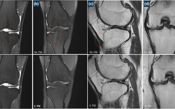
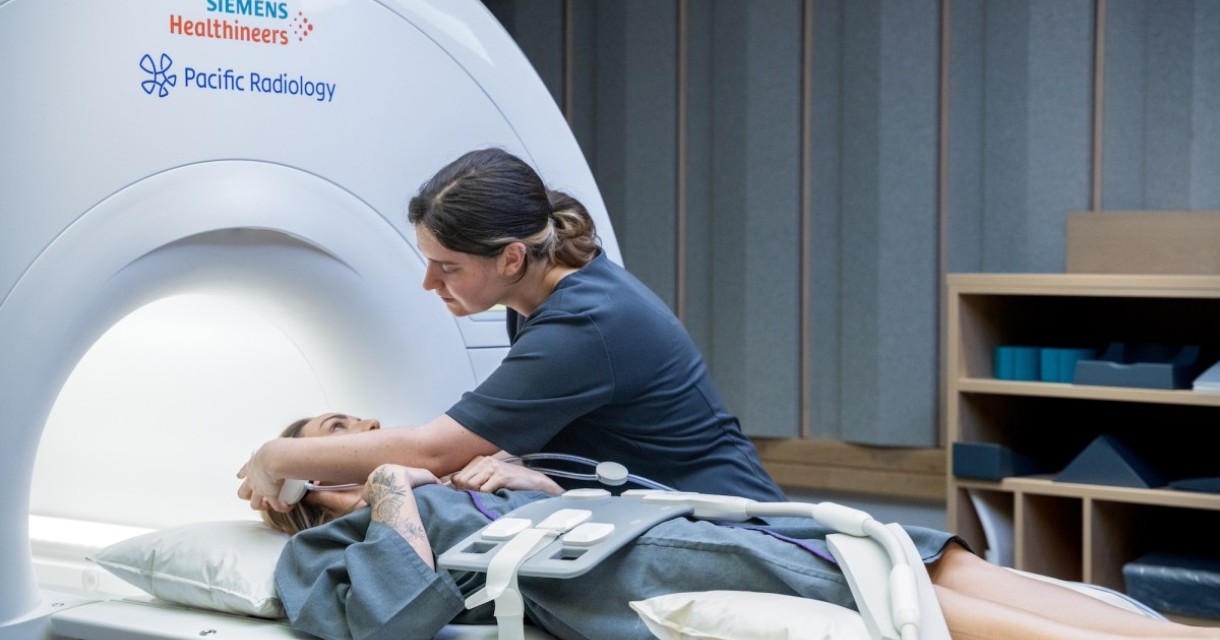
Pacific Radiology’s Research and Development Group has teamed up with leading German technology company Siemens, who also specialise in healthcare research and innovation. The study is beginning by investigating the improvements to knee scans, although the technology also has considerable implications for both cardiac and brain imaging. The team are currently in the clinical validation phase of the new technique and completing early phase assessment.
Dr Ross Keenan, Neuroradiologist and Head of Research and Development at Pacific Radiology, says the technology is truly ground-breaking, with potentially huge implications for patients and referrers:
"At Pacific Radiology we are committed to improving the patient journey. The efficiency of this new technology makes things much easier for our patients as they are in the scanner for the shortest possible time."
Deep Learning is a technique where radiologists can teach machines to recognise certain features in an image to identify what it is, using a method very similar to how humans learn. Using Deep Learning algorithms, we are then able to scan the knee much more efficiently than usual. Deep Learning is a technique that has started to evolve already in CT, and now Pacific Radiology is investigating the uses for MRI scanning. The idea is to provide much quicker scans because the machine knows what to look for, while producing images of equivalent or improved quality.
Benefits to this technology apart from faster scan times include superior resolution with incredibly fine detail, as well as the ability to remove ‘noise’ from images with a much purer signal. The team is even developing an anti-motion algorithm which can remove movement from patients in the scanner without having to repeat the scan. Considerably faster scan times also mean shorter wait lists for patients and referrers.
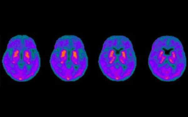
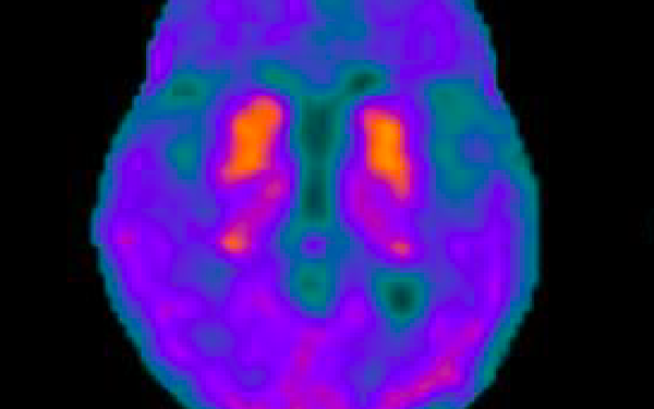
This technology also allows improvements which are beyond human limitations but extremely effective for machines, especially in cardiac and brain scanning. Radiologists can now easily perform strain imaging to measure stress on the wall of the heart, which was previously impossible. Radiologists have also taught the machine how to recognise white dots in the white matter of the brain, which is extremely useful in brain imaging, particularly to diagnose neurodegenerative disorders.
As Dr Keenan attests:
"This is really just the beginning of what could be an entire movement with potentially massive implications. If this learning algorithm works on the knee, we can perform quick scans with an equivalent or better image quality. We have even talked about using Deep Learning in PET scans in the future."
The study is progressing well, with 150 patients scanned so far and two specialist radiologists currently reviewing the data. The next phase of the study sees the team exploring the Deep Learning algorithm further by changing technical factors for more speed, more detail and better image quality, feeding back to Siemens in Germany.